


NCBI Isoforms:n |
crRNA Target Site:AGACTTCCTACATTGTGTCC |
Linker: LEADYKDDDDKRSEF |
Cas9:Wildtype spCas9 |
AICS-0063
DMD in WTC-mEGFP (mono-allelic tag)
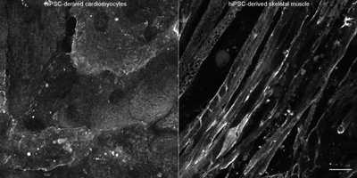
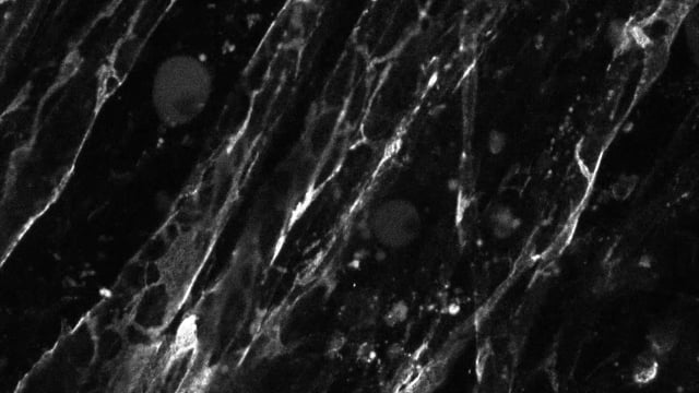
Single, mid-level planes of hiPSC-derived cardiomyocytes (left) and hiPSC-derived skeletal muscle myofibers (right) expressing mEGFP-tagged dystrophin imaged live in 3D on a spinning disk microscope. Left: Twelve days after the onset of differentiation, cells were plated on PEI and laminin coated glass and imaged 18 days later (30 days total after the onset of differentiation). Right: 25 days after the onset of differentiation, cells were plated on Matrigel (diluted 1:60) coated glass. Cell fusion was induced one day later, and muscle fibers were imaged 7 days after fusion induction (imaging was performed 33 days total after the onset of differentiation). Skeletal muscle sample was courtesy of Shawn Luttrell and David Mack (University of Washington). Brightness and contrast display settings are optimized for each panel; display settings are not the same between cell types. Scale bar, 20μm.

EditingDesign
GenomicCharacterization
StemCellCharacteristics
mEGFP Insert


