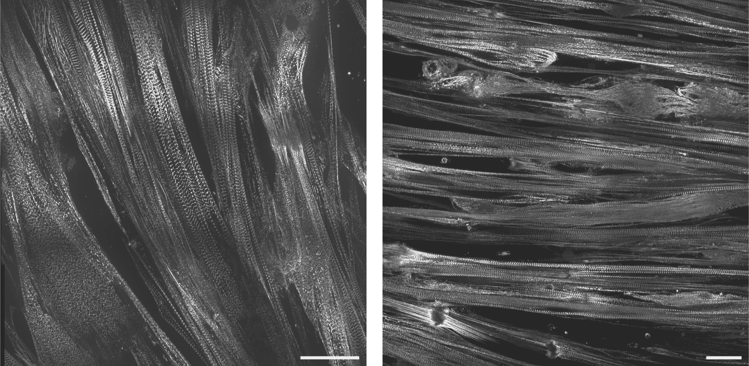
Live-cell imaging of skeletal muscle from the MYH3 G769V collection (Clone 82). Cells express mEGFP-tagged alpha-actinin-2. After 35 days of primary differentiation from hiPSC to myogenic progenitors, cells were replated onto Matrigel coated cover glass and induced to differentiate into skeletal muscle. Cells were imaged 11 days after this replating on a spinning disk confocal microscope. Images were acquired in a 3 x 3 tiled Z stack and are presented as maximum intensity projections of 10 slices. Scale bars are 50µm. Image system details: Nikon Eclipse Ti microscope with a Yokogawa CSU-W1 spinning disk head imaging onto an Andor iXon 888 EMCCD. Objectives were either Nikon Plan Apo VC 60x/1.4 NA or Nikon Plan Apo 100x/1.4 NA. Skeletal muscle sample and images was courtesy of Alina Greimal, BS, Christian Mandrycky, PhD and David Mack, PhD Institute for Stem Cell & Regenerative Medicine (ISCRM) at the University of Washington.