


crRNA Target Site:5’ GGAGATGGGTCCGCCCACCTGGG 3’ |
DNA Donor Sequence: Mutant* 5’ GAGGCAGAAGAGCCAGAGGAGATGGGTCCACCCACCTGGGCTCCTGAGCCGCTGGCAGA 3’ |
Cas9:TrueCut™ Cas9 Protein |
F Primer for PCR/ |
R Primer for PCR/ |
Red = PAM Site, Blue = Mutation
SNPNM_170707.4(LMNA):c.1824C>T(p.Gly608=) |
Gene SymbolLMNA |
Gene Namelamin A/C |
Parental LineAICS-0013 cl. 210 LMNB1 |
| Clone Number | Clone Type | Replicate | Genotype |
|---|---|---|---|
| 10 | Mutant | A | G608G/WT |
| 23 | Control | A | WT/WT |
| 39 | Mutant | A | G608G/WT |
| 45 | Control | A | WT/WT |

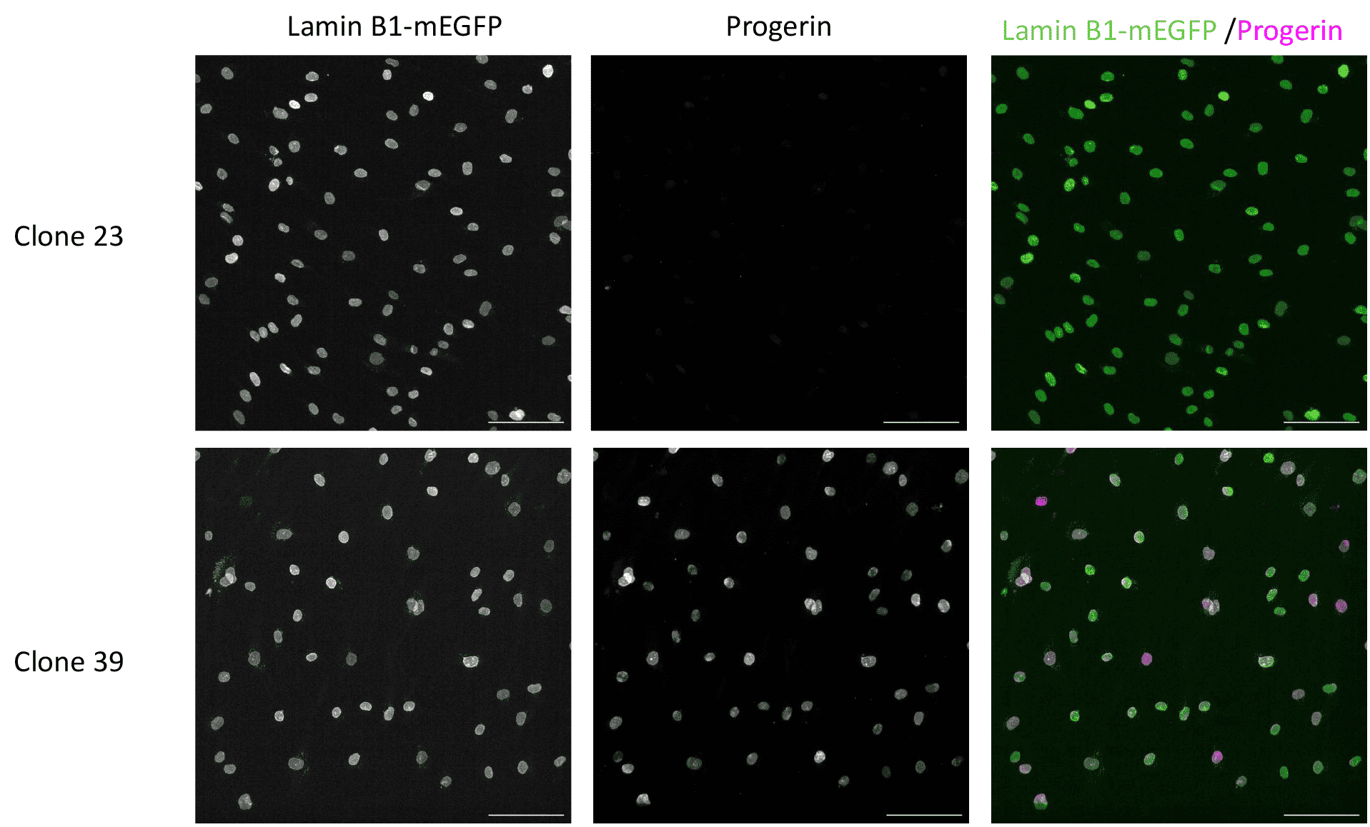
Immunofluorescent labeling of clones 23 and 39 to demonstrate expression of progerin proteins. hiPSCs were differentiated using the StemDIFF Endothelial Differentiation Kit (StemCell Technologies cat. No. 08005). Cells were cryopreserved in CryoStor CS10 (StemCell Technologies cat. No. 100-1061), and subsequently thawed into STEMdiff™ Endothelial Expansion Medium Kit (Catalog #08007) onto plastic tissue culture plates coated with Animal Component-Free Cell Attachment Substrate. Cells were passaged according to the provided protocol onto plastic tissue culture plates until Passage 7, at which point they were plated onto glass coated with Animal Component-Free Cell Attachment Substrate. After 4 days of culture the cells were fixed with a 4% paraformaldehyde in DPBS solution. Cells were then blocked and permeabilized in a solution of 1.5% normal goat serum + 0.4% Triton-100x in PBS for 1 hour at RT. Cells were labeled with an anti-progerin antibody (Santa Cruz cat. No. sc-81611, diluted 1:60) overnight at 4C. The following day cells were labeled with anti-mouse AlexaFluor594 (ThermoFisher cat. No. A-11005, diluted 1:500) for 1 hour at RT. Cells were imaged on a widefield microscope. All scale bars are 100um.


crRNA Target Site:5’ GGAGATGGGTCCGCCCACCTGGG 3’ |
DNA Donor Sequence: Mutant* 5’ GAGGCAGAAGAGCCAGAGGAGATGGGTCCACCCACCTGGGCTCCTGAGCCGCTGGCAGA 3’ |
Cas9:TrueCut™ Cas9 Protein |
F Primer for PCR/ |
R Primer for PCR/ |


